Beranda
/ Anatomy Rib Cage Muscles / Intercostal Muscles Anatomy A Schematic Picture Of Anatomy And Download Scientific Diagram - It is flexible and can expand and contract by the action of the muscles of respiration.
Anatomy Rib Cage Muscles / Intercostal Muscles Anatomy A Schematic Picture Of Anatomy And Download Scientific Diagram - It is flexible and can expand and contract by the action of the muscles of respiration.
Insurance Gas/Electricity Loans Mortgage Attorney Lawyer Donate Conference Call Degree Credit Treatment Software Classes Recovery Trading Rehab Hosting Transfer Cord Blood Claim compensation mesothelioma mesothelioma attorney Houston car accident lawyer moreno valley can you sue a doctor for wrong diagnosis doctorate in security top online doctoral programs in business educational leadership doctoral programs online car accident doctor atlanta car accident doctor atlanta accident attorney rancho Cucamonga truck accident attorney san Antonio ONLINE BUSINESS DEGREE PROGRAMS ACCREDITED online accredited psychology degree masters degree in human resources online public administration masters degree online bitcoin merchant account bitcoin merchant services compare car insurance auto insurance troy mi seo explanation digital marketing degree floridaseo company fitness showrooms stamfordct how to work more efficiently seowordpress tips meaning of seo what is an seo what does an seo do what seo stands for best seotips google seo advice seo steps, The secure cloud-based platform for smart service delivery. Safelink is used by legal, professional and financial services to protect sensitive information, accelerate business processes and increase productivity. Use Safelink to collaborate securely with clients, colleagues and external parties. Safelink has a menu of workspace types with advanced features for dispute resolution, running deals and customised client portal creation. All data is encrypted (at rest and in transit and you retain your own encryption keys. Our titan security framework ensures your data is secure and you even have the option to choose your own data location from Channel Islands, London (UK), Dublin (EU), Australia.
Anatomy Rib Cage Muscles / Intercostal Muscles Anatomy A Schematic Picture Of Anatomy And Download Scientific Diagram - It is flexible and can expand and contract by the action of the muscles of respiration.. The intercostals — the small muscles between your ribs — also contribute to the appearance of a muscular torso. The ribs are a set of twelve paired bones which form the protective 'cage' of the thorax. Each pair is numbered based on their attachment to the sternum, a bony process at the front of the rib cage which serves as an anchor point. These muscles attach the upper limb to the axial skeleton of the trunk and support the. As these muscles extend toward the front of the ribs, they become tapered, and at this point are referred to as the.
The transversus thoracic muscles originate from the posterior surface of the xiphoid process and the lower part of the body of the sternum. The ribs are attached to the breastbone, which is the. This muscle assists in depression of the ribs. So, let's learn the ribs so we can attach the muscles in the right place. Rib cage pain may be sharp, dull, or achy and felt at or below the chest or above the navel on either side.
2 4 2 Innermost Intercostal Muscles Proper from static.wixstatic.com The fibres pass superolaterally to insert into the internal surface of costal cartilages of ribs two to six. The intercostal muscles of the ribcage our ribcage exists to protect the heart and lungs. It is flexible and can expand and contract by the action of the muscles of respiration. According to the book clinical anatomy of the spine, intercostal muscles also influence the spine. These muscles attach the upper limb to the axial skeleton of the trunk and support the. The ribs are attached to the breastbone, which is the. The rib cage is collectively made up of long, curved individual. This muscle assists in depression of the ribs.
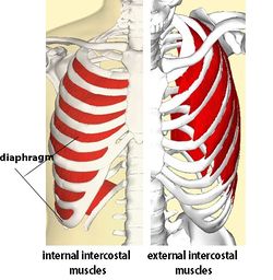
The muscles of the thoracic cage are the pectoralis major, pectoralis minor, serratus anterior, subclavius, intercostal (external, internal and innermost), subcostal and transversus thoracis muscles, including the diaphragm.
In this video, we explore:1) the anatomy of the sternum2) the anatomy and differences between the three classes of ribs3) the anatomy and differences between. It may occur after an obvious injury or without explanation. The transversus thoracic muscles originate from the posterior surface of the xiphoid process and the lower part of the body of the sternum. Start studying tx/rib cage anatomy. Labeled human stomach anatomy 6 photos of the labeled human stomach anatomy labeled human stomach anatomy, labeled human stomach anatomy picture, labeled human stomach diagram, stomach model labeled, stomach model labeled picture, stomach, labeled human stomach anatomy, labeled human stomach anatomy picture. The ribs are a set of twelve paired bones which form the protective 'cage' of the thorax. See more ideas about anatomy, anatomy study, rib cage anatomy. The direction of the fibres parallels that of the innermost intercostal. The rib cage intrinsically holds the muscles of respiration (diaphragm, intercostal muscles, etc.) that are crucial for active inhalation and forced exhalation, and therefore has a major ventilatory function in the respiratory system. These muscles attach the upper limb to the axial skeleton of the trunk and support the. The rib cage has three important functions: Diaphragm function in breathing what happens to it during exhalation: Related posts of rib cage diagram with organs labeled human stomach anatomy.
Rib cage pain may be sharp, dull, or achy and felt at or below the chest or above the navel on either side. But there's so many of them! Rib cage, in vertebrate anatomy, basketlike skeletal structure that forms the chest, or thorax, and is made up of the ribs and their corresponding attachments to the sternum (breastbone) and the vertebral column. This muscle assists in depression of the ribs. The external intercostal muscles consist of 11 muscles that envelop each side of the exterior of the rib cage from the back of the ribs and wrapping around where they are attached to the sternum in front.
Yoga For Spine Mobility Anatomy Of The Spine And Rib Cage Rib Cage Anatomy Yoga Fashion Rib Cage from i.pinimg.com These spaces are filled by intercostal muscles, and they also contain intercostal nerves and blood vessels. The external intercostal muscles or rib cage muscles (located between the ribs) contract together with the diaphragm, lifting and expanding the rib cage to provide even more space. Related posts of rib cage diagram with organs labeled human stomach anatomy. They comprise of thin slips of muscle, which run from the internal surface of one rib, to second and third ribs below. It makes up the thoracic wall, along with the skin, muscles, and fascia. The external intercostal muscles consist of 11 muscles that envelop each side of the exterior of the rib cage from the back of the ribs and wrapping around where they are attached to the sternum in front. Labeled human stomach anatomy 6 photos of the labeled human stomach anatomy labeled human stomach anatomy, labeled human stomach anatomy picture, labeled human stomach diagram, stomach model labeled, stomach model labeled picture, stomach, labeled human stomach anatomy, labeled human stomach anatomy picture. But there's so many of them!
The serratus ventralis thoracis muscle, attaching to the inner upper edge of the scapula and the side of the rib cage, forms a sling that supports the weight of the body.
The ribs are attached to the breastbone, which is the. It is flexible and can expand and contract by the action of the muscles of respiration. Start studying tx/rib cage anatomy. Rib cage, in vertebrate anatomy, basketlike skeletal structure that forms the chest, or thorax, and is made up of the ribs and their corresponding attachments to the sternum (breastbone) and the vertebral column. The thoracic cage presents with spaces between adjacent ribs, which are called intercostal spaces. This muscle assists in depression of the ribs. It makes up the thoracic wall, along with the skin, muscles, and fascia. It is made up of 12 pairs of ribs. Rib cage pain may be sharp, dull, or achy and felt at or below the chest or above the navel on either side. It may occur after an obvious injury or without explanation. As these muscles extend toward the front of the ribs, they become tapered, and at this point are referred to as the. It provides a strong framework onto which the muscles of the shoulder girdle, chest, upper abdomen and back can attach. The transversus thoracic muscles originate from the posterior surface of the xiphoid process and the lower part of the body of the sternum.
They comprise of thin slips of muscle, which run from the internal surface of one rib, to second and third ribs below. The intercostal muscles of the ribcage our ribcage exists to protect the heart and lungs. It provides a strong framework onto which the muscles of the shoulder girdle, chest, upper abdomen and back can attach. External intercostals muscle are the outermost layer lies directly under the skin originate from the lower border of rib above run obliquely and insert into the upper border of the rib below. It may occur after an obvious injury or without explanation.
Intercostal Muscle Strain Physiopedia from www.physio-pedia.com The external intercostal muscles or rib cage muscles (located between the ribs) contract together with the diaphragm, lifting and expanding the rib cage to provide even more space. Rib cage pain may be sharp, dull, or achy and felt at or below the chest or above the navel on either side. Start studying tx/rib cage anatomy. The intercostal muscles are those muscle bands that surround the ribs. The rib cage is an origin and insertion area for many muscles. The intercostals — the small muscles between your ribs — also contribute to the appearance of a muscular torso. The thoracic cage presents with spaces between adjacent ribs, which are called intercostal spaces. The ribs are a set of twelve paired bones which form the protective 'cage' of the thorax.
It may occur after an obvious injury or without explanation.
External intercostals muscle are the outermost layer lies directly under the skin originate from the lower border of rib above run obliquely and insert into the upper border of the rib below. It is made up of 12 pairs of ribs. The muscles of the thoracic cage are the pectoralis major, pectoralis minor, serratus anterior, subclavius, intercostal (external, internal and innermost), subcostal and transversus thoracis muscles, including the diaphragm. Anatomy the rib cage is a bony structure found in the chest (thoracic cavity). The rib cage is an origin and insertion area for many muscles. The external intercostal muscles or rib cage muscles (located between the ribs) contract together with the diaphragm, lifting and expanding the rib cage to provide even more space. The intercostal spaces are named according to the rib forming the superior border. Each pair is numbered based on their attachment to the sternum, a bony process at the front of the rib cage which serves as an anchor point. The intercostal muscles of the ribcage our ribcage exists to protect the heart and lungs. The rib cage is collectively made up of long, curved individual. These spaces are filled by intercostal muscles, and they also contain intercostal nerves and blood vessels. Learn vocabulary, terms, and more with flashcards, games, and other study tools. The serratus ventralis thoracis muscle, attaching to the inner upper edge of the scapula and the side of the rib cage, forms a sling that supports the weight of the body.
It may occur after an obvious injury or without explanation anatomy rib cage. According to the book clinical anatomy of the spine, intercostal muscles also influence the spine.